By age 44, endometriosis affects around one in nine women and people assigned female at birth in Australia.
It’s caused by the presence of tissue similar to the lining of the uterus found outside the uterus. While endometriosis is most commonly found in the pelvic cavity, it can sometimes be found in the diaphragm, lungs and elsewhere.
Symptoms include severe period pain, pain below the belly button when not menstruating, fatigue, digestive problems (often mistaken for irritable bowel syndrome), pain with bowel motions and/or urination, painful intercourse, and infertility.
It previously took, on average, 6.4-8 years for endometriosis to be diagnosed with surgery. But with doctors now able to give a clinical diagnosis of “suspected endometriosis” based on symptoms and a physical examination, the time to diagnosis is likely to reduce.
Read more: Endometriosis can end women's careers and stall their education. That's everyone's business
Why diagnose endo through surgery?
Endometriosis has historically been diagnosed through surgery. When performed by a skilled surgeon, this is still the most accurate method of diagnosis.
The most common surgical procedure for endometriosis is laparoscopy (or key-hole surgery). A thin telescope (called a laparoscope) is inserted into the belly button to see and access the organs inside the abdomen and pelvis.
Ideally, when the surgeon sees abnormal tissue during the procedure, they biopsy or remove a sample and send it to a lab. The pathologist then looks for endometrial-like cells under a microscope to provide confirmation of endometriosis. Occasionally, what a surgeon sees is not confirmed to be endometriosis but something else or normal tissue.

The endometriosis might be fully treated during that same diagnostic surgical procedure, or it might be incompletely treated or not treated at all. This depends on the extent of the endometriosis and the surgical skill of the surgeon, among other things.
Overall, surgery to remove endometriosis is effective in relieving pain symptoms, reducing infertility and improving quality of life.
However surgery is a very expensive way to achieve a diagnosis, both for the patient and the health system.
Laparoscopic surgery also comes with the risks of infection, major bleeding, and injury to important structures like the bowels or bladder. Recovery takes about four weeks.
How is the diagnostic process changing?
Some experts have argued surgery shouldn’t be used as a diagnostic test. This has prompted a move in recent years towards a “clinical diagnosis”, where a doctor makes an assessment based on symptoms and/or abnormal findings during a physical examination.
For most people, endometriosis symptoms begin with cyclical pain with their periods. That pain process evolves and pain can exist every day, with bowel motions or urination (often worse during the period), and during intercourse.
On physical examination, the doctor can sometimes feel endometriosis nodules in the vagina with the tips of their fingers. The lack of movement of the uterus as the doctor tries to move it with two hands may also raise suspicion, as can tenderness during this examination.
Read more: Considering surgery for endometriosis? Here's what you need to know
There are some drawbacks to clinical diagnosis. Most notably, the wrong diagnosis may lead a person down an incorrect treatment plan, inevitably delaying treatment for the true diagnosis.
People who receive a clinical diagnosis may also feel less able to access surgery, if that’s their preferred treatment, as a clinical diagnosis usually prioritises hormonal medications and other drug treatments in place of or before surgery.
Imaging techniques
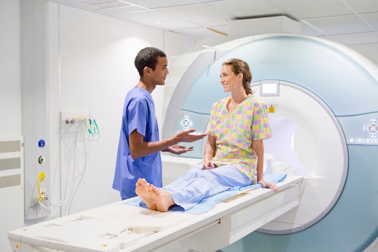
Over the past five to ten years, there has been an increasing ability to “see” endometriosis using imaging such as transvaginal ultrasound (an internal scan where the ultrasound wand is inserted into the vagina) and magnetic resonance imaging (MRI).
Diagnosing endometriosis through medical imaging is gaining popularity because it allows doctors and patients to understand the diagnosis and extent of the endometriosis without having to perform surgery.

The ability to see endometriosis relies heavily on the expertise of the person doing and interpreting the imaging test, just as seeing endometriosis at surgery relies on the expertise of the surgeon.
Not all types of endometriosis are yet reliably seen on an imaging test. For example, severe endometriosis with deep nodules and adhesions (bands of scarring which can attach to other organs) is easier to see than superficial endometriosis, which sometimes consists of a few deposits no larger than a few millimetres.
If the imaging is done by someone with expertise, it is generally possible to “rule out” moderate to severe endometriosis but minimal to mild disease may not be detected.
Read more: I have painful periods, could it be endometriosis?
Ideally, an imaging-based diagnosis should eliminate the need to have a two-step surgery (diagnostic surgery followed by treatment surgery), as the surgeon has a better understanding of the location and extent of the disease before starting the first surgery. This increases the likelihood of success with a single treatment surgery.
However, there are legitimate concerns that a move to use an imaging-based diagnosis will leave those with a “normal scan” falsely reassured because the disease is not visible on the scan. So, doctors should never tell someone they don’t have endometriosis based on an imaging test alone.
Mike Armour is the chair of the Endometriosis Australia research committee. He reports receiving funding from Metagenics, Canopy Growth, and Sci-Chem, outside the submitted work.
Cecilia Ng receives funding from the Medical Research Future Fund (MRFF).
ML reports receiving grant funding from OZWAC, Endometriosis Australia, AbbVie, CanSAGE, MRFF, HHS; honoraria for lectures/writing from GE Healthcare, Bayer, AbbVie, TerSera, consulting fees from Imagendo, outside the submitted work.
This article was originally published on The Conversation. Read the original article.


