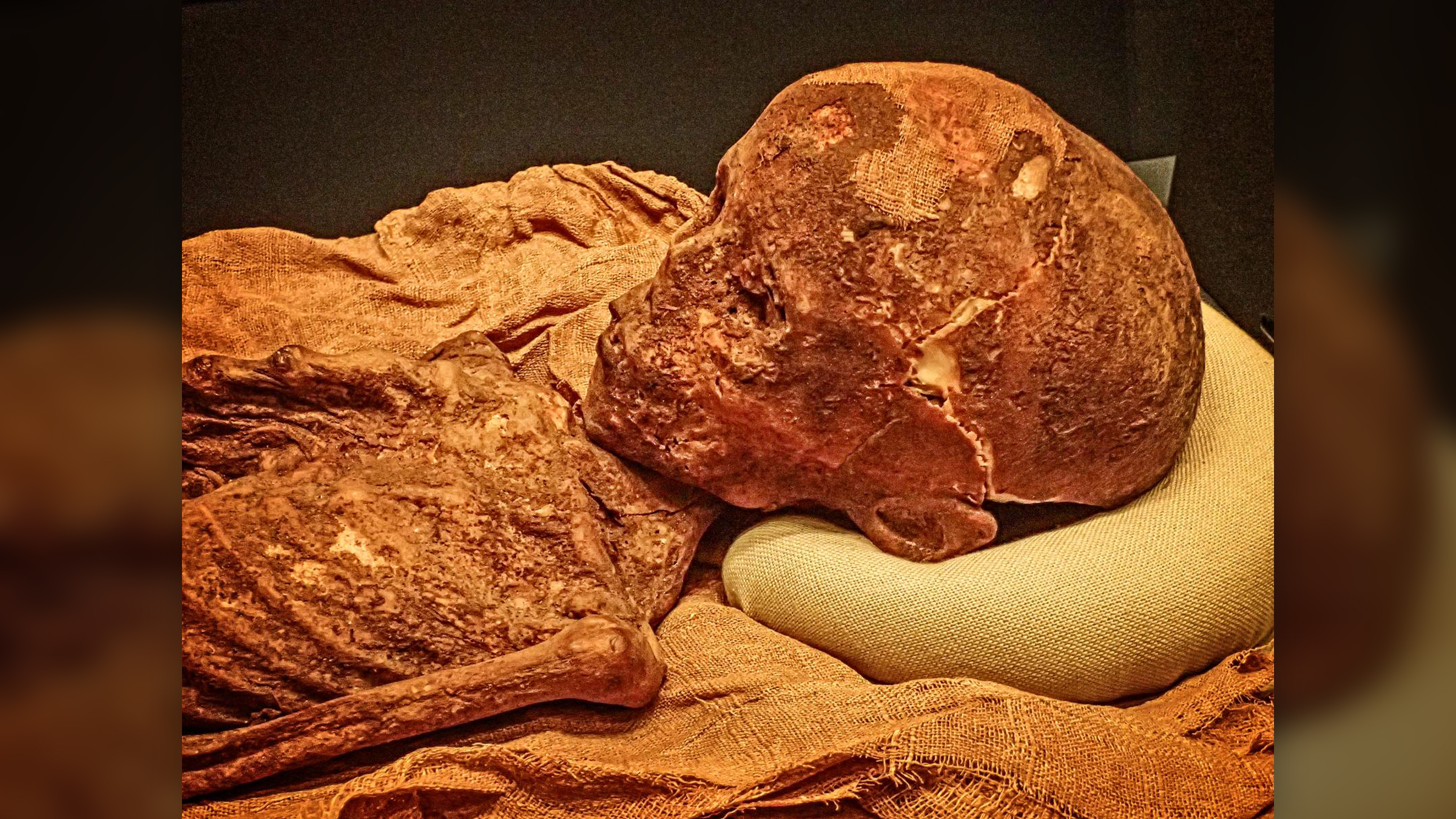
A large percentage of mummified children from ancient Egypt show signs of blood disorders known as anemias, suggesting that these youngsters may have had a slew of related medical problems, including malnutrition and growth defects, a new study finds.
Using full body CT (computed tomography) scans, a non-destructive way to study objects, an international team of researchers examined the remains of 21 child mummies who had died between the ages of 1 and 14. The team assessed the mummies for anemia by looking for telltale signs of the disorder, such as abnormal growth in the mummies' skulls and arm and leg bones.
Seven of the mummies, or 33% of those studied, showed signs of anemia in the form of thickened skull bones, the researchers found. Today, anemia is thought to affect 40% of children under the age of 5 years old globally, according to the World Health Organization.
This research on anemia in ancient Egypt "may shed light on ancient societies' health issues, dietary inadequacies, and social standards," Sahar Saleem, head and professor of radiology at Cairo University and a member of the Egyptian Mummy Project, told Live Science in an email. Saleem was not involved in the study.
Related: Little ancient Egyptian mummies hold surprises inside … and they aren't human
This study, published April 13 in the International Journal of Osteoarchaeology, is possibly the first of its kind to analyze the presence of anemia in mummified children. It includes child mummies from various parts of Egypt dating as early as the Old Kingdom (third millennium B.C.) to the Roman Period (fourth century A.D.).
Indigo Reeve, a bioarchaeologist at the University of Edinburgh in Scotland who was not involved in the study, defined anemia as "a lack of healthy red blood cells or hemoglobin." This condition can stem from a variety of causes, including dietary deficiencies, inherited disorders and infections, which can all lead to intestinal blood loss and poor absorption of nutrients, Reeve told Live Science in an email. Anemia typically causes fatigue and weakness, but it can also cause irregular heartbeat and can be life-threatening depending on the type and severity, she added.
Childhood cases of anemia can cause the expansion of some bone marrow, which is found at the center of most bones, which can lead to odd and abnormal bone growth, such as the thickening of the cranial vault, the part of the skull that holds the brain, Reeve explained. Porous lesions can also appear on bones, especially on the skull, which can cause further medical problems.
The study uncovered some of these anemia-related issues in the mummified children.
In one of the seven cases with thickened cranial vaults, a 1-year-old boy showed cranial signs of thalassemia, an inherited blood disorder that can cause mild to severe anemia due to reduced hemoglobin production; other symptoms of thalassemia can include inadequate and unusual bone growth and an increased risk of infections, according to Johns Hopkins Medicine. The boy also had an enlarged tongue and a condition known as "rodent facies," which is an abnormal growth of the cheekbones and an elongated skull. This boy's severe anemia, compounded with other difficulties, likely caused his death, the researchers theorized.
It's unclear how these ancient children came to have anemias, but the disorder can be caused by malnutrition, iron deficiency in pregnant mothers, chronic gastrointestinal issues and bacterial, viral or parasitic infections, all of which are thought to have been prevalent in ancient Egypt, the researchers said.
However, the study's small sample of 21 child mummies is not representative of an entire population or time period, the researchers noted. Further, the CT scans "produced blurry images due to low resolution that prevented interpretation" of additional signs of anemia, Saleem said.
"However," Saleem said, "we think that this work may pave the way for additional research on anemia and other ancient health issues in the future."


