What did the face of our ancestors look like 3 million years ago? Meet the reconstructed face of “Little Foot” – the most complete biological Australopithecus specimen that ever existed.
What did the face of one of our ancestors look like more than 3 million years ago? Our international team has answered this question by virtually reconstructing the facial fragments of Little Foot, the most complete Australopithecus skeleton yet discovered. This reconstruction sheds light on the influence of the environment on how our face evolved. Our findings have just been published in the Comptes Rendus Palevol journal, and the new 3D face of Little Foot can be explored online on the MorphoSource platform.
The search for human origins has never been more fruitful, with fossil discoveries pushing back the appearance of the earliest humans (members of the genus Homo) to 2.8 million years ago, and the development of cutting-edge methods for analysing these remains such as recovering genetic information from fossils over 2 million years old.
Yet, while our knowledge of extinct human species grows with each discovery, the story of our ancestors before the first humans appeared remains blurry. It is during this pivotal period that the traits defining our humanity emerged, enabling our genus’ evolutionary success.
Although the identity of our direct pre-Homo ancestor is far from resolved, one fossil group plays a central role in this search: Australopithecus. This genus, to which the famous “Lucy” belongs (discovered 50 years ago in Ethiopia), inhabited much of Africa and survived for over 2 million years. Australopithecus is known from many fossil remains, but often these are highly fragmentary, isolated, and have sometimes been distorted over the millions of years they have been buried. Notably, only a handful of skulls preserve nearly the entire face, a part of our anatomy that has profoundly shaped who we are today.
Through digestive, visual, respiratory, olfactory and non-verbal communication systems, the face is at the heart of interactions between individuals and their physical and social environments.
Significant changes occurred in the facial region throughout human evolution, with most structures generally becoming less robust. However, the factors driving these changes remain unclear. Were they caused by shifts in diet, social behaviour, or both? Only the discovery of more complete skulls can clarify this debate, and this is why the skull of Little Foot is crucial.
The ’Cradle of Humankind’
South Africa has been and remains a crucial region for researching into human origins. A century ago, the iconic “Taung Child” was published in Nature as a representative of a new African branch of humanity, Australopithecus. While scientific attention had previously focused on Eurasia, this discovery inspired decades of exploration and major finds across Africa. In particular, South Africa saw a proliferation of palaeontological sites in a region now UNESCO-listed and known as the “Cradle of Humankind.” Among these, Sterkfontein has proven exceptionally rich in fossils, many attributed to the hominin genus Australopithecus, and including numerous remarkably preserved specimens. But it was in 1994 and 1997 that Sterkfontein yielded its most spectacular find: the skeleton of Little Foot, over 90% complete, and the oldest human ancestor found in Southern Africa. To date, it is the most complete Australopithecus skeleton ever discovered, far surpassing Lucy, of which only 40% of the anatomy is preserved.
Our team has been studying this skeleton since its complete excavation concluded in 2017. The skull, in particular, has been the focus of our attention, as it is relatively complete, preserving all parts of the head – the cranium and mandible. However, 3.7 million years of burial underground have fragmented and displaced parts of its fossilised face. This process is especially visible in the forehead and eye sockets (orbits), making it impossible to quantitatively analyse these informative areas. Given the exceptional and unique nature of this fossil, we decided to harness the most recent technological advances in imaging to restore the face of Little Foot.
‘Little Foot’ in Europe
Creating a digital copy of Little Foot was essential to allow the virtual isolation and repositioning of the fragments without damaging the original skull. However, conventional X-ray scanning technologies have limitations. Through burial and fossilisation process, cavities were created in Little Foot’s skull as soft tissues disappeared and filled with sediment. As a result, X-rays struggle to penetrate this extremely dense sedimentary matrix, limiting image contrast and quality. After several unsuccessful attempts, we turned to a more powerful alternative: synchrotron radiation scanning. A synchrotron is a high-energy particle accelerator used to produce ultra-high-resolution images (at micron or even sub-micron scale).
With this in mind, we took Little Foot’s skull to England for scanning at the I12 beamline of the Diamond Light Source synchrotron. In the summer of 2019, Little Foot made its first journey outside Africa, carefully escorted across the world and housed in a secure vault during its stay in the UK.
A new face for Australopithecus
Several days were required to scan the entire skull at a resolution of 21 microns. The exceptional images generated revealed intimate details of Little Foot’s anatomy, and also provided the necessary data for facial reconstruction. However, the high quality of the data came at a computational cost: over 9,000 images were generated, representing terabytes of information to process. To virtually isolate the fragments, these images were processed using the supercomputer at the University of Cambridge (England). Once rendered in 3D, the fragments were repositioned according to their anatomical location, and missing parts were recreated to finally restore the complete face of Little Foot.
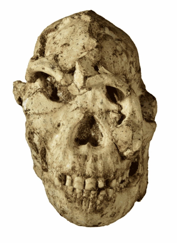
The size and shape of Little Foot’s orbits, previously obscured by displaced fragments, are among the most striking features of our reconstruction. In primates, the orbital region is heavily influenced by functional (visual) and behavioural (ecological) adaptations. Little Foot’s proportionally large orbits compared to other hominins suggest a strong reliance on sensory information, likely for foraging. This hypothesis is supported by a previous study showing that its visual cortex was more developed than that of modern humans.
The second major result of this study has implications for our understanding of the relationships between Australopithecus groups living in Africa between 4 and 2 million years ago. Although the comparative sample is limited, it includes specimens from both East and South Africa. Surprisingly, Little Foot, from a South African site, shows strong similarities with East African specimens. These similarities may indicate that Little Foot shared close ancestors with East African populations, while its probable descendants in South Africa later developed distinct anatomy through local evolution.
While the face provides valuable insights into our ancestors’ adaptations to their environment, the rest of Little Foot’s skull will offer further key elements for understanding our evolutionary history. Notably, the braincase, affected by “plastic” deformation, will require similar work to reconstruct and explore the neurological features of this fossil group.
This research was supported by the Agence Nationale de la Recherche, the Centre National de la Recherche Scientifique, the Claude Leon Foundation, the DST-NRF Center of Excellence in Palaeosciences, the French Institute of South Africa, Diamond Light Source, and the ISIS facility of the Science and Technology Facilities Council (STFC).
Dominic Stratford est membre de organisation: Department of Anatomical Sciences, Renaissance School of Medicine, Stony Brook University, Stony Brook, NY, 11794, USA Funding: Already stated by Dr Beaudet
This article was originally published on The Conversation. Read the original article.