Get all your news in one place.
100's of premium titles.
One app.
Start reading
LiveScience
LiveScience
Emily Cooke

3D map plots human brain-cell 'antennae' in exquisite detail

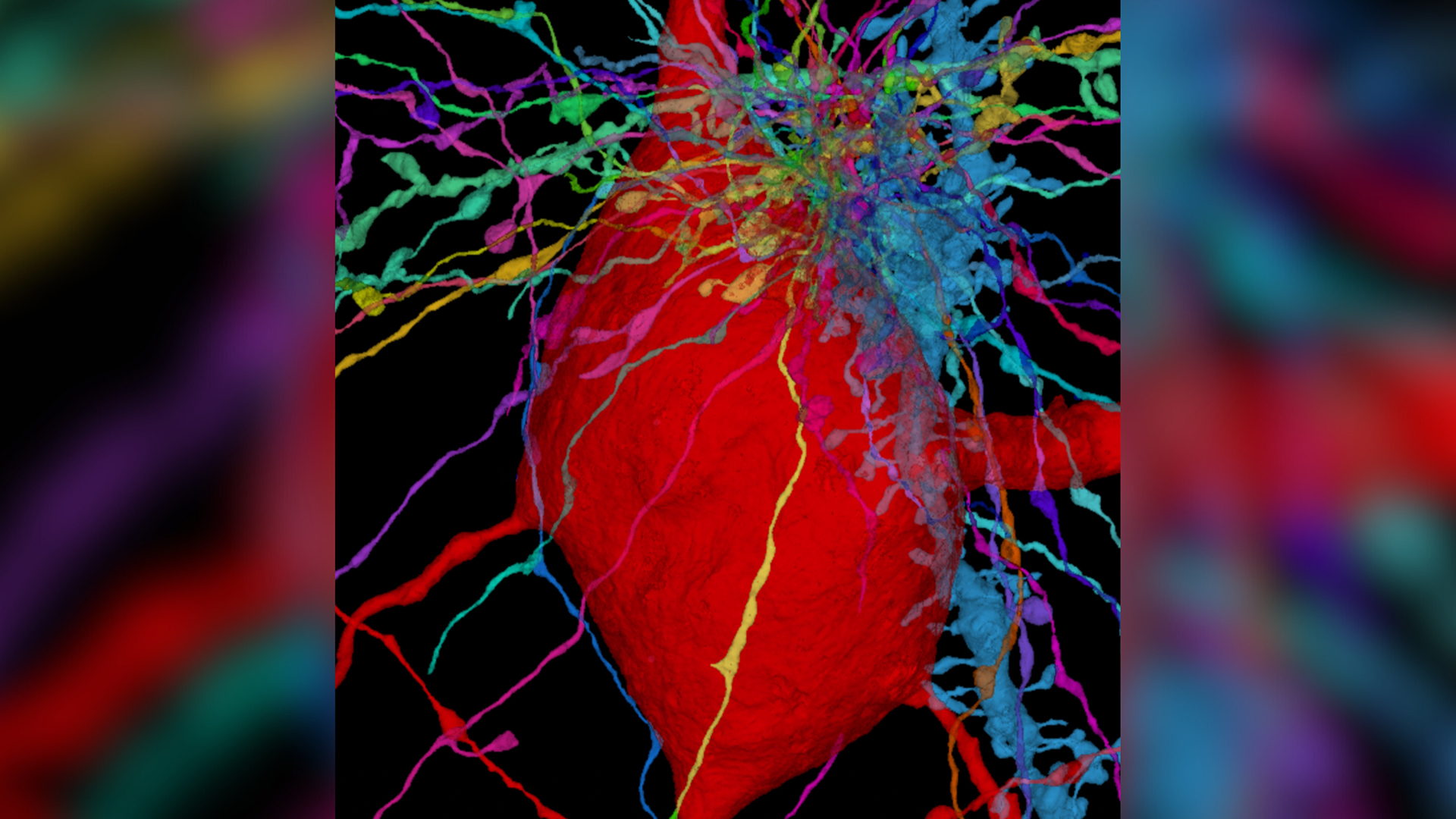
This microscope image shows the "wires" extending from neurons (multicolor) interacting with a specialized type of neuron called an interneuron (red) in the human cerebral cortex. The interneuron has a hairlike antenna called the primary cilium (also red) that's hidden within the meshwork in the upper-right corner.

Tiny, hairlike "antennae" protrude from the surface of brain cells, and now, scientists have unveiled a detailed map of these wires across the whole human cortex. They hope the new map will guide future research into a class of diseases that cause these structures to malfunction.

The hairlike structures, known as cilia, are actually found on the surface of most eukaryotic cells, meaning complex cells that house their DNA in a nucleus. Some cilia can move; for example, cilia in the lungs collectively beat to clear the airways of harmful pathogens. Others, called primary cilia, are immobile and instead act like antennae, sensing signals from their environment and passing them to the nucleus of the cell.

Sign up to read this article
Read news from 100's of titles, curated specifically for you.
Already a member? Sign in here
Related Stories
Top stories on inkl right now
One subscription that gives you access to news from hundreds of sites
Already a member? Sign in here
Our Picks
Fourteen days free
Download the app
One app. One membership.
100+ trusted global sources.